
pain bump on back of heel
Painful Bump on the Back of Your Heel: How to Choose the Right Next Step
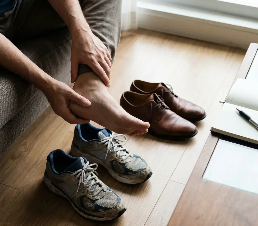
Start by Matching the Bump to the Pain
A visible bump and heel pain can occur together without the bump being the pain source. For readers in Tustin, California, a useful starting point is to compare the pain’s exact location with what touches it, when symptoms began, and whether rest or activity changes them. This is a way to organize the symptoms, not a way to name the condition.
The painful structure may be a bony prominence, the Achilles tendon, a nearby bursa, irritated soft tissue, or another source of heel pain. These possibilities can overlap. Pressure from a rigid shoe, loading around the Achilles region, and pain under the heel point to different questions that still require clinical context.
Treat the visible contour and the painful area as two observations. If they line up where a shoe presses, contact may be relevant; if pain follows the Achilles or sits under the heel, the bump may not explain it. That comparison can guide what to record and discuss without establishing a diagnosis.
Before deciding what the bump may involve, ask whether the pain and the visible prominence occupy the same place. Pain where a rigid shoe presses raises a shoe-contact question; pain along the Achilles region after rest or loading raises a question about the Achilles or nearby tissue; pain under the heel raises a different set of possible causes. These observations organize the next question but do not diagnose the source.
Keep a separate urgency screen. Sharp or sudden heel or Achilles pain, visible swelling, or difficulty walking warrants prompt medical evaluation. If none of those warning signs is present, note whether symptoms track direct pressure on the bump, Achilles loading, or another location. Take that distinction to an evaluation if the pattern persists, recurs, or limits activity.
Match the Pain’s Location and Timing
Start by matching the tender area to the visible bump. Pain directly where a rigid shoe contacts the back of the heel keeps a bony prominence or irritated nearby tissue in the question. Pain along the Achilles insertion raises a tendon or bursa question, while pain under the heel shifts attention toward plantar heel pain and its different possible causes. A bump with tenderness somewhere else may represent two findings rather than one source.
Heel pain can have local, referred, or systemic causes. Reported local causes include plantar fasciitis, heel fat-pad atrophy, calcaneal stress fracture, and nerve entrapment. Referred causes and systemic illnesses are also possible. This range is why the location of the pain matters more than the visibility of the bump alone.
Record the exact painful area rather than only describing the prominence. Note whether tenderness is behind the heel, at the tendon’s attachment, below the heel, along the sides, or in more than one place. The comparison gives an evaluation a clearer source-matching question without turning the observation into a diagnosis.
Pain timing adds another clue. Pain at the back of the heel that is worse after rest can occur with Haglund’s syndrome, but that pattern does not confirm the condition. Record whether the first steps after sitting or sleeping are uncomfortable, whether activity increases symptoms, and whether the pain eases or returns after movement. The sequence matters because timing is part of the history a clinician uses to sort possible sources.
Shoe contact is another practical observation. A rigid shoe counter that presses on the bump may irritate nearby tissue when a bony enlargement is present. You can note whether sandals, soft-backed shoes, or shoes with a different heel height change the pressure. A change in pressure may help you identify an aggravating factor; it does not prove what the bump is.
Also notice whether the pain is present at rest or mainly appears with walking, running, climbing, or another activity. A gradual pattern after increased loading raises a different question from pain that began at a specific moment. Neither pattern supplies a diagnosis, but both are useful details to report.
Your short symptom record can include:
- The exact spot where pain begins: behind the heel, under it, along the tendon, or elsewhere.
- Whether the bump is on one side or both sides.
- Whether symptoms began gradually or after a clear event.
- Whether rest, walking, exercise, or a particular shoe changes the pain.
- Whether swelling, altered walking, or reduced activity has appeared.
- Whether the discomfort is present at rest, after rest, during activity, or afterward.
- Whether the pattern has remained stable, returned repeatedly, or changed over time.
Bring those observations to an evaluation if the symptoms do not settle. They give the discussion more detail without turning your notes into a self-diagnosis.

A Bony Prominence May Irritate Nearby Tissue
Haglund’s deformity describes an enlargement of the bony heel near the Achilles insertion. The enlarged bone can irritate nearby soft tissue when it rubs against rigid shoes. This explanation fits a pattern in which the bump is located at the back of the heel and pressure from the shoe counter increases discomfort, but the pattern still does not identify the bump with certainty.
The word “deformity” describes a structural finding; it does not tell you how much pain you should have or whether the finding is responsible for every symptom. A visible prominence can exist alongside another source of pain. That is why the relationship between the bump, the tender area, footwear, rest, and walking deserves attention.
Pressure can matter even when the shoe is not painful during every step. Repeated contact may irritate tissue over time, while a change in shoe shape may reduce contact without changing the underlying structure. Treat that response as information about an aggravating factor, not as confirmation of Haglund’s deformity.
The cause of Haglund’s deformity is not well known. Suggested contributors include a tight Achilles tendon, a high arch, and heredity. These are possible contributors, not a checklist that lets you identify the cause from your own foot shape or family history.
You may notice that both heels have a similar contour, while only one side hurts. You may also notice that pain is more pronounced after rest or when a rigid shoe presses the area. Those observations can help frame the clinical question, yet they cannot determine whether the bony prominence, tendon, bursa, or another structure is producing the pain.
Do not assume that a family pattern, a high arch, or tightness explains the entire problem. Those features are discussed as suggested contributors, while the overall cause remains uncertain. The relevant question is how the finding relates to your symptoms and function.
For suspected Haglund’s syndrome, clinical evaluation and lateral ankle radiographs are often sufficient for diagnosis. That statement applies to a suspected condition after assessment; it does not mean every painful bump requires an X-ray or that an image alone explains your symptoms. If pressure from footwear is a clear aggravating factor and no warning sign is present, you can use the pressure-reducing measures described later while deciding whether the problem is settling or needs evaluation.
Achilles and Bursal Symptoms Can Overlap With a Bump
Achilles tendon and bursal problems can create pain in the same general region as a posterior-heel bump. Achilles tendinopathy is associated with excessive mechanical loads on the gastrocnemius and soleus muscles. That mechanical-loading explanation is useful, but the causes of Achilles tendinopathy are not fully established. A guideline notes ongoing uncertainty about its causes, diagnostic criteria, imaging role, natural course, treatment choices, long-term prognosis, and recurrence prevention.
This distinction matters because a bump does not automatically mean that the tendon is the source of pain, and tendon pain does not automatically mean that the bump is the cause. Your activity history, symptom timing, tenderness, function, and examination findings all affect the question.
A gradual increase in activity may be part of the history when tendon loading is being considered, but the absence of an obvious activity change does not rule out other explanations. Keep the description broad until an assessment connects the symptoms with a particular structure.
Retrocalcaneal bursitis is another possible contributor to pain near the back of the heel. If retrocalcaneal bursitis is involved, lifting the heel and loosening the shoe counter may help control pain. The conditional wording matters: those measures are described for a relevant bursal pattern, not as proof that a bursa is involved in your case.
The same review describes heel lifts, rocker shoes, and the AirHeel Brace as options that can reduce Achilles tendinopathy symptoms. These options belong to a broader care discussion rather than a universal instruction. A shoe change that reduces pressure may be reasonable to observe when your symptoms are mild and there are no warning signs, while persistent or function-limiting pain calls for assessment.
A response to a heel lift or altered shoe does not distinguish reliably between tendon, bursal, and bony causes. Several structures can respond to a change in pressure or load. Note what changes, but avoid treating that change as a test result.
You can organize the overlap by asking three questions:
- Does the pain seem tied to direct pressure from the shoe counter?
- Does the problem relate more to loading the Achilles region during activity?
- Is there a visible bump, swelling, or change in walking that needs examination rather than observation?
These questions sort the next conversation; they do not supply a diagnosis. If the answer is unclear, do not force the symptoms into a tendon, bursa, or bone category before an evaluation.
If more than one pattern appears at once, describe each part separately. For example, you can report pressure from a shoe, pain after activity, and tenderness at the back of the heel without deciding that all three have one cause.

Pain Under the Heel May Point to a Different Question
Pain under the heel may point to a different question from pain at the back of the heel. Plantar heel pain can arise from local conditions such as plantar fasciitis, heel fat-pad atrophy, calcaneal stress fracture, or nerve entrapment. Referred causes and systemic illnesses can also produce heel pain. A posterior bump may be noticeable at the same time without being the source of pain under the heel.
Use the location of your most meaningful pain rather than the most noticeable finding. If the bump is visible but the tenderness is mainly on the underside of the heel, describe both facts separately. That distinction prevents the visible bump from becoming an assumption about the cause.
Pain under the heel can also coexist with posterior-heel discomfort. In that situation, reporting two pain locations is more useful than choosing one explanation too early. The location, timing, and effect on walking help determine whether the assessment should consider more than one structure.
The range of possible plantar heel causes also explains why different evaluations may consider different questions. History and physical examination are important for identifying the pattern. Depending on the concern, clinical assessment may involve bone, soft tissue, nerves, or a broader health context.
Do not treat an unusual location, a recurring symptom, or a second pain site as a reason to keep trying the same self-care measure indefinitely. Use those details to decide whether you need a more complete assessment.
A bump at the back of the heel should not make you ignore pain under the heel, and pain under the heel should not automatically be attributed to the bump. Describe both locations, especially if one is changing how you walk or limiting ordinary activity.
Separate a Pressure Problem From a Sudden Injury Pattern
Sharp or sudden heel or Achilles pain, difficulty walking, or visible swelling warrant prompt medical evaluation. Those symptoms change the decision from watching the bump to seeking assessment. Do not wait for the bump’s appearance to become clearer before responding to a sudden change in pain or function.
A sudden onset can be especially meaningful when symptoms follow an event, a change in activity, or a forceful movement. You do not need to determine whether the tendon, bone, bursa, or another structure is involved before arranging prompt care. Describe what happened, when the pain began, whether swelling appeared, and what walking now feels like.
The key issue is the change in severity or function, not whether the visible bump looks dramatic. A sudden change deserves attention even if the skin and shape appear familiar.
Use this simple boundary: mild, stable symptoms without warning signs can be observed with general pressure-reducing measures, while sharp or sudden pain, visible swelling, or difficulty walking calls for prompt medical evaluation. If your symptoms are severe or changing quickly, prioritize timely clinical attention over experimenting with footwear, stretching, or activity.
When you seek care, lead with the change that triggered concern. Say whether the pain was sudden, whether walking became difficult, and whether swelling appeared. Those details help communicate why observation is no longer the appropriate first step.
When the Pattern Outranks the Appearance
Red flags are clinical prediction guides associated with a higher risk of serious disorders such as infection, inflammation, cancer, or fracture. Red flags are not diagnostic tests, and they do not by themselves establish a diagnosis or prognosis. Their purpose is to raise the level of attention and help determine whether assessment should happen promptly.
For a painful heel bump, that means you should not use a familiar explanation such as shoe rubbing or a possible bony prominence to dismiss a concerning change. A symptom that looks ordinary can still need evaluation when the overall pattern raises concern.
A red flag does not tell you which serious condition is present. It tells you that self-diagnosis is not an adequate basis for waiting. Let the concern determine the urgency of assessment rather than trying to prove or disprove a specific label at home.
Test Whether Contact Is Part of the Pattern
When no warning sign is present, general measures can help clarify whether contact or loading is contributing, but they cannot identify the bump. Conservative options for rearfoot problems can include shoe modification, insoles, or orthoses; for a Haglund’s deformity, reducing shoe contact with the lesion may alleviate pain. General heel-pain measures described in the sources include padding or heel cups, avoiding flat shoes and barefoot walking, limiting extended high-impact activity, and cryotherapy. These are general options, not a personal plan.
Keep the observation focused on the source question. If a softer or differently shaped shoe counter changes pain at the prominence, record that relationship. If the pain remains under the heel or along the Achilles, or if a change increases symptoms, that pattern is more useful to bring to an evaluation than to interpret as proof of what the bump is.
You can keep the observation period practical:
- Choose footwear that reduces direct contact with the back of the heel.
- Consider padding, a heel cup, an arch support, or an orthotic insole as described options.
- Avoid prolonged barefoot walking and flat shoes if those conditions aggravate the heel.
- Limit extended high-impact activity while you observe how the symptoms respond.
- Apply cryotherapy to the affected area as described for general heel-pain care.
- Note whether walking, pressure, or rest-related pain changes.
Stop treating observation as the right approach if symptoms become sudden, swollen, difficult to walk with, persistent, recurrent, or activity-limiting. A short record of what you tried and what changed can make the next evaluation more focused.
Initial heel-pain treatment options may include padding and strapping, orthotic insoles, oral anti-inflammatories, or a corticosteroid injection selected and administered by a clinician. These options should not be interpreted as a personal treatment plan for a painful bump. Medication and injection choices depend on the clinical question and the person being evaluated, so keep those decisions in the assessment setting rather than adding them to self-care on your own.
Stretching also deserves restraint. General guidance describes Achilles and plantar fascia stretching, but a painful bump with sudden symptoms, swelling, or difficulty walking should be assessed rather than pushed through a stretching routine.
A measure described for one type of heel pain may not address another. Do not assume that a treatment mentioned for plantar heel pain is appropriate for posterior-heel pain, or that an option discussed for a tendon problem applies to a bony prominence.
The purpose of general measures is limited: reduce avoidable pressure, observe the symptom pattern, and decide whether the problem is settling or needs evaluation. They are not a way to confirm Haglund’s deformity, Achilles tendinopathy, bursitis, plantar heel pain, or any other condition.
When the Bump and Pain Stay Out of Step
Persistence matters when the bump, the painful area, and function do not move together. If the prominence remains visible but pain stays under the heel, follows the Achilles, returns with activity, or changes how you walk, do not assume that the bump explains every symptom. Arrange a clinical evaluation when symptoms persist, recur, worsen, or limit activity.
The course of a defined Achilles condition cannot be transferred automatically to an unexplained bump. A time point or rehabilitation experience from one condition does not establish what is happening at a different location. Persistent or recurrent symptoms should be assessed in context, especially when reducing shoe contact does not change the painful area.
Bring a record of the prominence’s location, direct shoe contact, pain location, rest and activity triggers, and walking changes. That information helps an evaluation address whether the visible bump and painful tissue correspond.
Most people with Achilles tendinopathy respond well to nonsurgical care such as activity modification, physical therapy, eccentric exercises, and tendon-loading programs. Recovery can still require commitment to rehabilitation and may take time. For midportion, noninsertional Achilles tendinopathy, the cited source reports approximately 89% success rates with eccentric exercise protocols.
That reported figure applies to a defined Achilles tendinopathy pattern and a specific exercise protocol. It does not apply automatically to insertional pain, a visible heel bump, plantar heel pain, a bursal problem, or an unexplained symptom. Do not transfer a result from one diagnosis to another simply because both involve the heel.
If a clinician identifies a tendon-related problem, the rehabilitation plan may depend on the tendon location, symptoms, activity demands, and examination findings. A general exercise result cannot select that plan for you.
A small percentage of people have persistent Achilles tendinopathy symptoms after conservative treatment lasting several months to more than 1 year, and surgery may then be considered. This longer time frame shows why persistence needs context: some tendon problems require extended rehabilitation, while sudden or function-limiting symptoms may need attention immediately.
Arrange evaluation when the pain does not improve, repeatedly returns, worsens, interferes with walking or activity, or no longer fits the pattern you were observing. Bring your timeline, footwear changes, activity changes, and any response to general measures.
Do not use a long rehabilitation timeline to justify waiting through a sudden change. The expected course for a defined tendon condition is different from the decision to seek prompt assessment after sudden pain, swelling, or difficulty walking.
Before an appointment, write down:
- How long the bump and pain have been present.
- Whether symptoms are improving, stable, recurring, or worsening.
- Which activities you have stopped or changed.
- Whether footwear or pressure changes the symptoms.
- Whether you have had swelling, sudden pain, or difficulty walking.
- What general measures you tried and what happened afterward.
This record helps you explain why observation is no longer enough and keeps the evaluation focused on your actual pattern.
Use Examination and Imaging to Match the Source
An evaluation can focus on whether the visible prominence and the painful tissue correspond. The history may cover when each began, whether a shoe counter presses on the area, whether pain is worse after rest or activity, whether symptoms are under the heel or along the Achilles region, and whether walking changed. The examination may compare the prominence, Achilles region, nearby soft tissue, function, and the painful side with the other side.
Ultrasound can evaluate the Achilles tendon for tears and tendinopathy and assess nearby bursas, with performance depending in part on the operator and equipment. A lateral ankle radiograph addresses a different question by showing bone and a suspected bony prominence. The selected test depends on which structure or relationship needs clarification.
Ask which structure the clinician is evaluating and whether the visible bump appears to correspond to the painful tissue. That keeps the discussion centered on the bump-to-pain relationship without predicting a diagnosis or treatment plan.
Imaging may be considered when the clinician needs to examine bone, tendon, or nearby bursas. Lateral ankle radiographs can help assess a suspected bony prominence.
An imaging choice follows the clinical question. A picture of a bony prominence may not explain soft-tissue pain, and an assessment of the Achilles does not answer every question about pain under the heel. Ask what structure the proposed test is intended to assess and how the result would affect the next discussion.
Imaging is therefore one part of clarification, not a substitute for describing the symptom pattern. The useful question is not simply whether a test can be performed, but what unresolved concern it is meant to address.
Useful questions for an evaluation include:
- Which area appears to be producing the pain: the bone, tendon, bursa, plantar tissues, nerve, or another source?
- Does the location and timing fit the working explanation?
- Is imaging needed to answer a specific unresolved question?
- What finding would change the recommended activity or footwear guidance?
- If symptoms persist, which type of follow-up is appropriate?
- Does the visible bump appear related to the painful tissue, or could the two findings be separate?
These questions help you understand the reasoning without asking for a diagnosis from symptoms alone. Bring your symptom record so the discussion includes timing, triggers, walking, and changes in footwear rather than only the visible bump.
Let the Bump-to-Pain Match Guide the Next Step
Use the bump-to-pain match to choose the next step. If pain is centered where a shoe contacts the prominence, remains mild and stable, and no warning sign is present, general contact-reducing measures can provide a cautious observation period. If pain is under the heel, follows the Achilles, stays separate from the bump, or persists, recurs, worsens, or limits activity, arrange an evaluation rather than assuming the prominence is the cause. Sudden pain, visible swelling, or difficulty walking warrants prompt medical evaluation.
At an appointment, bring the comparison that matters: where the pain is, what touches it, how rest and activity affect it, and whether walking changed. Ask which structure is being assessed and whether the bump appears to correspond to the painful tissue. The goal is to clarify that relationship, not to assign a label from appearance alone.
A qualified clinician should decide your individual suitability for examination, imaging, activity guidance, medication, injection, rehabilitation, or any other treatment after considering your symptoms and health history. Until then, describe the bump by its location, timing, triggers, shoe contact, and effect on walking. That information gives your evaluation a clear starting point without assuming what the bump means.
Questions to Bring to a Heel-Pain Consultation
Is a pain bump on the back of the heel always Haglund’s deformity?
No. A visible prominence may involve bone, irritated Achilles or bursal tissue, shoe pressure, plantar heel pain, injury, or another local, referred, or systemic cause. Location, timing, footwear contact, activity, swelling, and walking changes can guide an evaluation, but symptoms alone cannot establish the cause.
What should I do if the lump or pain appeared suddenly?
Sudden or sharp heel or Achilles pain, visible swelling, or difficulty walking warrants prompt medical evaluation. Do not try to identify the structure or test it with stretching, exercise, or footwear changes before seeking appropriate care, particularly if the symptoms followed an injury or forceful movement.
Can changing shoes reduce pain from a posterior-heel bump?
It may reduce irritation when a shoe counter presses on the area. Shoes that limit direct contact, along with options such as padding, heel cups, insoles, or orthoses, can be general conservative measures for mild, stable symptoms without warning signs. Improvement with footwear does not confirm the cause or remove the underlying prominence.
When should persistent heel-bump symptoms be evaluated?
Arrange an evaluation when discomfort persists, returns with activity, worsens, changes how you walk, or limits normal activity. A clinician may use the history and examination to decide whether imaging is useful; lateral radiographs can help assess a suspected bony prominence, while ultrasound can evaluate the Achilles tendon and nearby bursas. The appropriate test depends on the unresolved clinical question.
Free consultation
Find out whether regenerative treatment is right for you.
Most consultations begin as a telehealth call with a physician specialist, so you can start without travelling to a clinic. No referral needed.