
what are bone spurs in the heel
What a Heel Bone Spur Means—and What It Cannot Tell You About Heel Pain
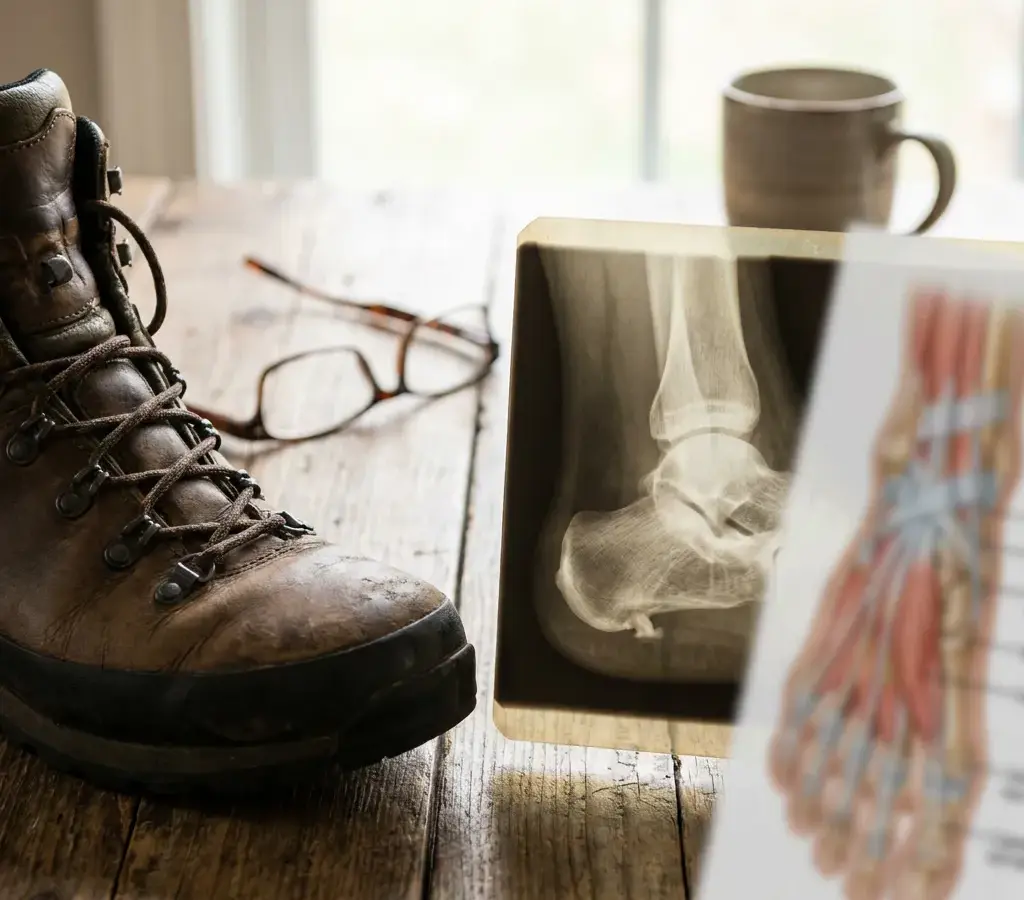
What a plantar calcaneal spur describes
A heel spur is a bony projection on the heel bone, also called the calcaneus. A spur on the bottom of the heel is described as a plantar calcaneal spur, an outgrowth from the heel bone. “Plantar” identifies the sole-facing side of the foot, and “calcaneal” refers to the calcaneus, or heel bone. The longer phrase makes the location of the bony projection explicit.
One historical explanation tied plantar spurs to traction from a tight plantar fascia. Another proposal points to weight-bearing compression with secondary traction. Calcaneal spurs have been reported in 10% to 63% of people without symptoms. The theories describe candidate processes, but a visible outgrowth does not identify which one produced it. The reported range describes occurrence, not which proposed pathway led to the outgrowth.
Plantar fasciitis is the most common cause of plantar heel pain, and a spur can be present with or without that condition. Those are separate pieces of information: one describes a bone finding, while the other describes a possible source of pain. The useful question is not only whether a spur appears on the image, but how it fits with the symptoms you are experiencing.
The phrase “heel spur” can sound like a complete explanation because it names a specific structure. It is more limited than that. The finding tells you that extra bone is present at the heel; it does not, on its own, establish when or why it formed, whether it is related to your pain, or whether another condition better explains your symptoms.
Several theories may explain how heel spurs form
The formation of a plantar calcaneal spur does not have one settled explanation. Proposed theories include degenerative change, inflammation, traction, repetitive trauma, bone-forming tendencies, and vertical compression. These are possible explanations, not a confirmed sequence that applies to every spur. The terms refer to different proposed influences: degenerative change and bone-forming tendencies concern changes involving bone; inflammation concerns an inflammatory response; traction describes pulling; repetitive trauma involves repeated stress; and vertical compression refers to pressure through the heel. Some anatomical descriptions locate the outgrowth at the origins of the flexor digitorum brevis and abductor digiti minimi muscles rather than within the plantar fascia itself. This anatomical distinction helps explain why a simple fascia-traction account remains debated: if the outgrowth is described at nearby muscle origins rather than within the fascia, the fascia is not necessarily the structure at the spur’s attachment site. That does not rule out traction as one proposed influence, but it cautions against treating it as a settled explanation. A vertical orientation has also been discussed in relation to compression rather than a horizontal pulling force. One proposal combines weight-bearing compression with secondary traction. These accounts distinguish pulling at a tissue attachment from pressure through the heel, though one proposed route allows both forces to be involved. They illustrate why the cause remains contentious rather than establishing one route for all spurs. A review also identifies reported associations with age, weight, gender, arthritides, plantar fasciitis, and foot position as possible factors in spur formation. These observations broaden the discussion beyond a single local pulling force, but an association does not prove that any one factor caused an outgrowth. The review also notes discrepancies in descriptions of spur anatomy, histology, and clinical associations. These are separate uncertainties: descriptions of where a spur is located, its tissue characteristics, and how it relates to symptoms do not by themselves establish what initiated its growth. The proposed mechanisms remain candidate explanations, not a history that can be inferred for an individual spur.
One proposed pathway involves the plantar fascia, the band of tissue attached near the heel. Repetitive microtrauma or traction involving the fascia may lead to inflammation, which can trigger new bone formation where the tissue attaches to the bone. This describes one possible way a spur might develop; it does not establish that every spur formed this way.
The direction of the relationship between a spur and plantar fasciitis remains uncertain. A spur might develop after inflammation, or the two findings might be connected in another way. Other proposed explanations include vertical compression and degenerative change. Taken together, these theories do not provide a reliable account of what caused a particular person’s spur.
For you, the practical point is that a formation theory is not a diagnosis of your pain. The possibility that repeated irritation plays a part does not establish that you have plantar fasciitis, nor does it show that the spur itself hurts. A proposed mechanism can describe how an outgrowth may arise; it cannot, by itself, explain your symptoms.

Plantar fascia problems and spurs can overlap without proving cause
Heel spurs and plantar fasciitis can appear together, but overlap does not show which one caused your symptoms. Heel spurs are present in approximately 50% of patients with plantar fasciitis. That figure measures how often the findings coexist in that group; it does not mean the spur causes half of those patients’ pain. Its denominator matters: it starts with people who have plantar fasciitis and reports how often a spur is also present. It does not answer the inverse question of how many people with a spur have plantar fasciitis, or how much the spur contributes to pain. The reported 10% lifetime prevalence of plantar fasciitis in the general population measures a different thing; it is not a measure of spur-related pain. These statistics concern different populations and questions, so they cannot be turned into an individual probability or a causal estimate.
The presence of a spur does not correlate well with plantar fasciitis symptoms, and spurs also occur in people without plantar fasciitis. A spur on an X-ray therefore cannot tell you, by itself, whether plantar fasciitis explains your heel pain. The image and your symptoms provide different kinds of information, and the first does not settle the second.
Plantar fasciitis often involves throbbing pain along the inner-bottom part of the heel that is worse with the first steps after rest. The pain may ease after you have walked for a while, then return with continued weight-bearing. These features describe a pattern associated with plantar fasciitis; they do not make an X-ray spur proof of the cause of your pain.
Plantar fasciitis is the most common cause of plantar heel pain, but its cause is often unclear and may involve more than one factor. Possible risk factors include obesity and work that involves prolonged standing and weight-bearing. Those points concern plantar fasciitis; they do not establish why a particular spur formed or prove that the condition explains your pain.
Keep three observations separate: a spur is visible, plantar fasciitis may or may not be present, and your heel hurts.
Reported associations do not settle a spur’s origin
A plantar calcaneal spur may be an indicator of foot pain even apart from plantar fasciitis, without necessarily causing the pain itself. It may point to other associated conditions. That distinction matters because a finding can be clinically associated without revealing a biological origin. The study describes a plantar calcaneal spur as a possible indicator of foot pain independent of plantar fasciitis, while noting that the spur may not cause the pain and may indicate other associated conditions. The word “indicator” describes the reported relationship; it is not an account of how the bone formed. Formation theories address proposed forces or biological processes, while a clinical association describes findings observed together. Neither type of evidence automatically resolves the other question: a reported relationship with pain does not identify whether traction, inflammation, compression, or another proposed process contributed to the spur. Nor does a proposed formation pathway show that a spur is a direct pain source. Keeping these questions distinct lets a possible association remain relevant without turning it into proof of cause or a settled explanation of formation. The cited study reports a possible indicator and associated conditions, not a mechanism that can be assigned from the finding alone.
Heel spurs are rarely the direct cause of heel pain, although they can cause symptoms if they press on nerves or other tissues in the foot. That possibility is different from assuming that a spur shown on your X-ray is pressing on something or causing your symptoms. The image alone does not establish that connection.
A useful way to interpret the finding is to leave room for more than one possibility: the spur may be incidental, or it may be associated with a condition relevant to your pain. Neither an association nor the possibility of pressure on nearby tissue proves that the spur is causing your symptoms. The finding deserves context, not an automatic verdict.

Persistent or worsening heel pain merits professional evaluation
Persistent or worsening heel pain should not be ignored. Midwest Foot & Ankle Center advises seeking medical attention for this kind of pain in its guidance for patients in Fort Dodge and Humboldt, Iowa. If your pain continues or gets worse, arrange a professional evaluation rather than treating the spur as a complete explanation.
An evaluation can focus on the pain as well as the image: what might explain your symptoms, and what does the spur add to that picture? A plain side-view X-ray showing a spur does not by itself diagnose plantar heel pain. Since spurs can occur with or without plantar fasciitis, the image needs to be considered alongside the problem you are trying to understand.
Your description of the pain provides a separate kind of information from the X-ray. In plantar fasciitis, pain often affects the inner-bottom part of the heel, is worse with the first steps after rest, and may return with continued weight-bearing. You can note whether any of those features match your experience and explain what does not. A pattern can help frame the question, but it does not establish a diagnosis on its own. An examination may add observations that a radiograph cannot provide. In plantar fasciitis, pressing the medial calcaneal tuberosity and the plantar fascia typically produces sharp, stabbing pain, and passive dorsiflexion of the foot and toes often elicits pain. These are reported features of plantar fasciitis, not proof that a visible spur caused the pain. Pressure tenderness and movement-related pain are distinct observations: one concerns response to touch around the heel attachment, while the other concerns symptoms when the foot and toes are moved upward. They add a physical-examination perspective alongside your description of when pain occurs and the image.
An X-ray finding and a symptom history answer different questions. Imaging is not required to diagnose plantar fasciitis, though weight-bearing radiography, MRI, or ultrasound can help rule out other causes of heel pain. Seeing a spur therefore does not make it the explanation by default; the relevant question is what the image contributes to understanding your symptoms.
A short note can help you describe why you are seeking attention. Include how long the pain has persisted, whether it is worsening, what the X-ray report calls the finding, and whether pain is worse with the first steps after rest or returns with continued weight-bearing. Use the note to explain your concern, not to diagnose yourself. The spur on the report remains one piece of information, not a complete answer. Heel pain can also interfere with walking, exercise, work, or other daily activities. If it affects you in these ways, describing which activities are affected adds practical context about the problem you want evaluated, without suggesting what caused it.
Use an evaluation to understand what the finding means for you
A heel spur describes a bone finding; it does not, by itself, explain your heel pain. Spurs can coexist with plantar fasciitis, appear without symptoms, or be associated with foot pain without directly causing it. If your pain persists or worsens, seek professional attention rather than assuming the spur tells the whole story. A qualified professional can assess what the finding means in your individual situation.
Free consultation
Find out whether regenerative treatment is right for you.
Most consultations begin as a telehealth call with a physician specialist, so you can start without travelling to a clinic. No referral needed.