
pain on top of the feet
Pain on the Top of the Feet: How to Choose Your Next Step

Start with what top-of-foot pain can—and cannot—tell you
Pain on the top of the feet is a symptom, not a diagnosis. It can have overuse, structural, or medical causes; less common possibilities include tarsal coalition and ganglion cysts. These categories are broad starting points rather than conclusions. The location, timing, triggers, associated symptoms, and direction of change help organize the questions that need answering, but symptoms alone cannot establish which explanation applies. Use the pattern to decide what information to describe and whether temporary observation or clinical clarification makes sense. The goal is to identify a reasonable next action without turning a familiar symptom into a label.
Your next step depends on four practical details: where the pain is concentrated, what changed before it began, whether the pain followed an injury, and whether warning signs are appearing. Walking ability, swelling, skin changes, altered sensation, and the direction of symptoms over time add context. Symptoms alone cannot reliably distinguish a fracture from a bruise or sprain, so a reassuring guess should not replace an evaluation when the pattern remains unexplained.
Use this guide as a sorting process rather than a home diagnostic test. First map the symptom. Then connect its timing to activity, footwear, or an injury. Next look for changes that alter the urgency. Finally, decide whether temporary observation is reasonable or whether you need an examination. The goal is a clearer action, not a label. This sequence also guards against common shortcuts. A familiar location does not identify a particular tissue, and a recent activity or footwear change does not prove that it caused the pain. Being able to walk does not reliably exclude a fracture, while pain without one clear accident does not exclude a stress fracture. Gather the pattern first, screen for changes that alter urgency, and use the result to choose a question and a next step—not to turn clues into a diagnosis.
A clinical evaluation can bring together your history, a physical examination, and, when appropriate, foot X-rays. That matters because two people can describe pain in a similar place while needing different questions answered. Keep your attention on the pattern you can describe and the changes that should move you toward care.
Map the painful area to possible structures
The exact location of top-of-foot pain gives your evaluation a starting question, although it does not establish the cause. Pain over the middle of the foot can arise from arthritis in the small midfoot bones. Pain in that region may also involve anterior tibial tendonitis, an overuse problem affecting a tendon that helps lift the foot. Pain over the tendons on top of the foot can fit an extensor tendon problem when excess pressure or strain has been present. Those location clues are most useful when paired with the symptom’s context. For example, discomfort centered in the midfoot that is aggravated by tight lacing gives the clinician a joint-related question to consider, because midfoot arthritis may worsen with shoe pressure. A midfoot symptom appearing after repeated activity can also make an overuse tendon question relevant, including the anterior tibial tendon, which helps lift the foot. Pain over the top tendons after added pressure or strain creates a different discussion from pain accompanied by fatigue and muscle cramps, which can occur with tarsal coalition. None of these pairings confirms a cause. Their value is in preventing an overly broad description such as “my foot hurts” and showing which structure, trigger, or associated symptom should be examined.
A structural possibility can look different from an overuse pattern. Tarsal coalition means that two or more middle foot bones are abnormally fused, and it can cause pain, fatigue, or muscle cramps. Ganglion cysts are another less common possibility identified in discussions of top-of-foot pain. These examples are prompts for a clinical examination, not labels for you to apply from location alone.
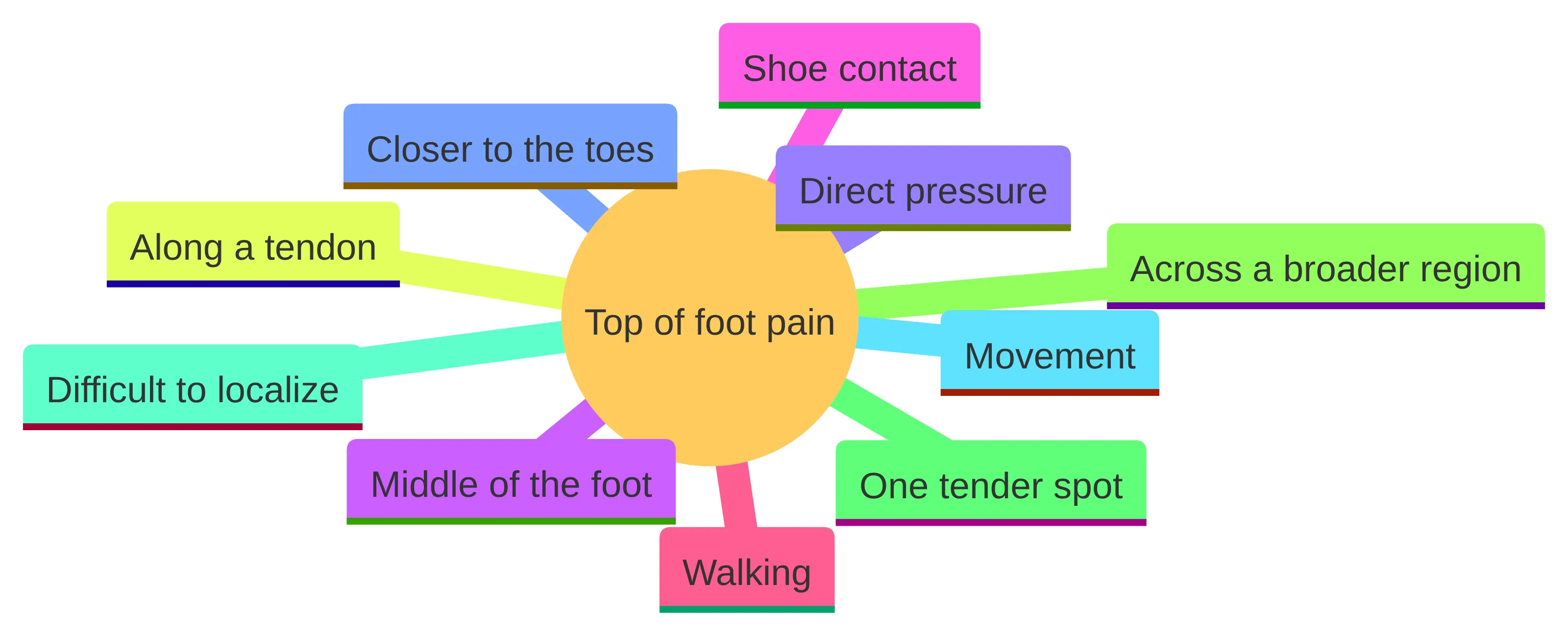
Make the map more precise by noting whether the discomfort is close to the toes, centered over the midfoot, near the ankle, or spread across the upper surface. Record whether you can point to one tender spot or whether the area is difficult to localize. Also note whether movement, direct pressure, shoe contact, or walking changes the symptom. These distinctions help you explain the problem without turning a location into a diagnosis.
Start your notes by marking the most precise area you can: the middle of the foot, closer to the toes, along a tendon, or across a broader region. Record whether the same spot hurts when you press it, move the foot, put on a shoe, or walk. The purpose is to describe the pattern clearly, not to prove which structure is responsible.
Use activity and footwear changes as context, not proof
A recent change in activity can explain why the timing deserves attention, but it cannot identify the cause by itself. Stress fractures are small cracks in bone caused by repeated stress rather than one sudden injury. A sudden increase in exercise intensity or a change in training surface can contribute to that stress, while repeated pressure or strain on the tendons on top of the foot can contribute to extensor tendonitis.
Footwear can also change the pattern you notice. Pain from arthritis in the small midfoot bones may worsen when shoes are tied too tightly. If the pain predictably changes with lacing, shoe pressure, walking, running, or a new training routine, record that contrast. A useful comparison is not simply whether a change occurred, but whether the timing lines up. Note the last activity or footwear change before symptoms, whether the pain was present at rest, and whether it returned when the same demand was repeated. In a runner or other high-impact athlete, an increase in intensity or a new surface matters because repeated forces affect weight-bearing bones; it still does not establish a stress fracture. Likewise, pressure on the top of the foot can be relevant to an extensor tendon problem, while tight lacing may aggravate pain associated with midfoot arthritis. Treat these observations as questions for evaluation, not as a way to select treatment from a guessed diagnosis. A trigger is useful context; it is not proof that footwear, exercise, or a tendon is the entire explanation.
Write down what changed and when. A new running route, a longer walk, more time standing, a different training surface, or tighter lacing may be relevant even if the change seemed minor. Include the activity level you tolerated before the pain and the activity that first made it noticeable. This timeline is more useful than a general statement that you have been “more active.”
Do not use an activity increase as a reason to push through worsening pain to see what happens. When a stress fracture is possible, continuing activity can worsen the injury and delay recovery. A recent training change therefore raises a question about evaluation rather than giving you permission to keep testing the foot.
Separate pain after an injury from pain without one
Pain after an injury calls for a different first screen than pain that developed without one. Note the event, the foot position, the force involved, and what changed immediately afterward. Inability to bear weight after an injury warrants prompt evaluation, and a severe injury may require emergency care. A new deformity or suspected broken bone also needs prompt medical attention.
Pain without one clear accident does not rule out a bone problem. Repeated loading can produce a stress fracture, particularly in weight-bearing bones of the feet and lower legs. Runners and people in high-impact sports have greater risk, and a sudden increase in exercise intensity or a change in training surface can add stress. If persistent pain follows that kind of change, arrange an evaluation rather than assuming that no fall means no injury. That distinction matters because a stress fracture is a small crack produced by repeated stress, not necessarily by one dramatic accident. The relevant history may therefore be cumulative: training intensity, surface, and weight-bearing demand over time. A person may remember only a gradual increase in pain. The absence of a single event does not remove the need to describe the timeline or the possibility that imaging may be useful. Since symptoms can overlap with a bruise or sprain, use the history to identify the question and an examination to resolve it.
Being able to walk does not rule out a fracture because some broken bones still allow weight-bearing. If walking is possible but pain persists, worsens, or becomes more localized, treat walking ability as one observation rather than a clearance signal. Use the trend and associated symptoms to decide how quickly to seek care.
For an injury-related symptom, record whether the pain began immediately or developed later. Note swelling, bruising, a change in shape, difficulty taking steps, and any numbness or unusual temperature in the toes. For pain without a clear injury, record recent loading and whether the pain keeps returning with activity. The two histories may lead to different examination priorities.
Burning, tingling, or numbness changes the clinical question
Burning, tingling, numbness, or unusually sensitive skin shifts the question toward possible nerve involvement rather than pain from a tendon or joint alone. Sural nerve compression or trauma may cause neuropathic pain, burning, or increased skin sensitivity in the foot and nearby lower leg. The common peroneal nerve supplies movement and sensation to the lower leg, foot, and toes; dysfunction of that nerve is a form of peripheral neuropathy. The type of sensory change and any movement change provide different information to report. The sural nerve is primarily sensory and follows a pathway through the lower leg to the foot; compression or trauma may cause burning or heightened skin sensitivity in the foot and nearby lower leg. Because the common peroneal nerve is involved in both movement and sensation, report any movement change alongside numbness or tingling. This comparison does not identify which nerve is involved; it helps you avoid collapsing every altered sensation into the same explanation.
Describe the sensory change precisely. Record whether it is burning, pins and needles, reduced feeling, or pain from light contact; whether it stays in one area or travels; and whether it appears with walking, at rest, or after an injury. Include any change in movement or toe control. A nerve-related pattern still does not identify the exact nerve or cause from symptoms alone, but it gives a clinician a more useful starting point.
New numbness or tingling after an injury needs prompt evaluation, especially when the toes also look pale or bluish or feel unusually cold. Those changes may signal a nerve or circulation problem. Do not wait for a sensory change to become severe before treating it as a reason to seek prompt care.
Avoid treating a burning or tingling sensation as proof of neuropathy, nerve entrapment, or a circulation problem. Use the symptom quality to decide what to mention and how quickly to act when it follows an injury or occurs with toe color or temperature changes. A clinician can then determine which possibilities require attention.
Watch how the symptom changes over time
The direction of top-of-foot pain matters more than a single snapshot. Note whether the pain is easing, staying at the same level, returning with the same activity, spreading, or becoming more severe. Severe or worsening pain, substantial swelling, or bruising may require medical assessment. Persistent pain after a change in activity also deserves attention because continuing activity with a stress fracture can worsen the injury and delay recovery. Trend changes the question in a way a single pain score cannot. Improvement after the suspected trigger stops may be useful context, but it does not prove that the trigger was the cause. A stable symptom still deserves a record if it keeps returning with the same load. Worsening pain, new swelling, or bruising may require medical assessment, while a new sensory or skin change changes the warning-sign screen. Use the trend to decide whether observation is still temporary or whether the pattern now needs clinical clarification.
Use a simple time record rather than relying on memory. Write down when the pain began, what you were doing before it began, what makes it better or worse, and whether walking or shoe pressure changes it. Add any swelling, bruising, skin change, burning, tingling, numbness, or difficulty bearing weight. This record helps you recognize a worsening pattern and gives a clinician a clearer account.
Compare the symptom with ordinary activity without deliberately provoking it. Ask whether a short period of normal movement leaves the pain unchanged, increases it, or brings it back later. Record how long the change lasts. Do not turn repeated walking, running, jumping, or pressing on the painful area into a test of whether the foot is injured.
A symptom that has not settled or that remains unexplained should move you from observation toward evaluation. There is no need to wait for the pain to become extreme before arranging care. The purpose of tracking is to clarify the next decision, not to create a home diagnosis.
Stop monitoring when urgent warning signs appear
An open wound requires urgent care. Severe bleeding or bone visible through the skin calls for emergency evaluation. Spreading redness, warmth, drainage, or fever can indicate infection and warrants prompt medical assessment. Do not treat those signs as ordinary soreness from activity or footwear.
After an injury, new numbness, tingling, pale or bluish toes, or unusual coldness needs prompt evaluation because it may signal a nerve or circulation problem. If you have diabetes, seek prompt evaluation for a foot injury or wound because reduced sensation and slower healing can increase risk. These warning signs change the timing of care even when the pain itself is not the strongest symptom.
Use the warning signs as stop points, not as a checklist for proving what happened. You do not need to identify a fracture, infection, nerve problem, or circulation problem before seeking care. Describe the change, when it began, and whether it followed an injury. The presence of a warning sign is enough to change your next action.
When no warning sign is present, keep monitoring temporary and general
When no warning sign is present, monitoring should be temporary and used to collect information, not to confirm a diagnosis. Keep a brief record of pain location, timing, activity, footwear pressure, walking ability, and associated symptoms. If pain clearly worsens with an activity, do not keep repeating that activity as a test; continued activity can worsen an undiagnosed stress fracture and delay recovery.
Use the lowest-risk general boundary while you observe the pattern: avoid sudden increases in exercise intensity and changes in training surface, and notice whether tight shoe lacing aggravates the pain. These details are associated with possible causes, but changing them does not prove that the pain will resolve or that you have identified the source. Do not add medication, devices, or exercises based on a guessed diagnosis.
A temporary monitoring plan should answer three questions: Is the pain changing? Is ordinary walking becoming harder? Has any warning sign appeared? Check those questions at reasonable intervals rather than repeatedly pressing or loading the painful area. Write down the answer and the activity around it so you can see whether the pattern is stable, recurring, or worsening.
Stop monitoring and arrange care if the pain persists, worsens, returns repeatedly, or remains unexplained. Seek prompt attention sooner if any injury, wound, infection sign, circulation change, or new sensory change appears. A temporary observation period has a clear boundary: it should not delay evaluation when the pattern is no longer stable. The absence of a warning sign should be read narrowly. It does not confirm that the pain is a bruise, tendon problem, or uncomplicated overuse injury, because symptoms alone cannot reliably distinguish a fracture from a bruise or sprain. If the symptom remains unexplained, the purpose of monitoring has been reached: you have collected context to share with a clinician, not a result that rules out another cause.
Persistent or unexplained pain merits clinical evaluation
Persistent foot pain deserves an evaluation even when no single injury explains it. A podiatrist can evaluate persistent foot or ankle pain for a possible stress fracture, with imaging used to help confirm the diagnosis when needed. More generally, evaluation may include a physical examination, questions about your symptoms, and foot X-rays; the appropriate treatment depends on the cause and severity. Each part of that process answers a different kind of uncertainty. Your history supplies the timeline, activity and footwear context, and associated changes. The physical examination lets the clinician assess the painful area and relate what you report to the foot’s findings. Foot X-rays may be used when appropriate, and imaging can help confirm a suspected stress fracture when needed. Because the same top-of-foot location can involve a joint, tendon, bone, nerve, or structural issue, an evaluation is how the next question is selected before treatment is discussed. Ask what the clinician is trying to distinguish and what finding would change the plan.
Unexplained pain also merits evaluation because several categories can overlap. Midfoot arthritis, tendon problems, stress injury, nerve compression, and structural conditions can produce different combinations of pain, activity sensitivity, swelling, fatigue, burning, or altered skin sensitivity. A clinical assessment can sort those questions in a way that symptom matching at home cannot.
Arrange care when your notes show a persistent or recurring pattern, when the pain limits ordinary walking, or when you cannot explain why it began. Persistent does not have to mean unbearable. Repeated disruption of walking or activity is enough to make clarification useful, particularly when the pattern keeps returning after temporary improvement.
Bring the pattern rather than a preferred diagnosis. Explain where the pain is, what changed before it began, what makes it worse or better, and which associated symptoms have appeared. The goal of the appointment is to clarify what needs attention before discussing treatment.
Bring a clear symptom record to the evaluation
A useful top-of-foot pain record can include:
- The exact location and whether one or both feet are affected.
- The date or approximate period when the pain began.
- Any fall, twist, impact, new exercise, mileage change, training-surface change, or other activity change before it began.
- Whether shoe pressure or tight lacing changes the pain.
- Whether walking is possible and what happens during or after activity.
- Swelling, bruising, redness, warmth, drainage, fever, or a visible change in shape.
- Burning, tingling, numbness, unusual sensitivity, pale or bluish toes, or unusual coldness.
- Whether the pattern is improving, stable, recurring, or worsening.
The record gives a clinician specific questions to ask during the history and examination. You can also bring a list of relevant health conditions, previous foot injuries, and care already tried, along with the change you noticed after each one. Keep the description factual: “pain increased after a new running surface” is more useful than “I have a stress fracture.” Add any prior evaluation, imaging, or treatment and what happened afterward, but do not treat a response—or lack of response—as proof of the cause. If one or both feet hurt, say whether the onset, location, and triggers match or differ. This comparison gives the clinician a clearer starting point for deciding which part of the history and examination needs closer attention.
Use the appointment to ask what findings support each possible explanation, whether imaging is appropriate, and what change would require faster follow-up. If a stress fracture is part of the question, ask how the examination and any imaging address it. These questions keep the discussion focused on the source of pain and the next decision rather than on a treatment chosen in advance.
Keep the record short enough to use during the visit. A dated list or phone note can work: symptom, activity, footwear, associated change, and what happened next. Mark the first day you noticed the pain, the point at which it changed, and any period when it returned after improving. If both feet hurt, describe whether the pattern is similar on each side or different. Also separate the symptom’s starting pattern from later changes: note whether pain became more localized, began occurring at rest, or was joined by swelling, bruising, altered sensation, or difficulty bearing weight. This sequence helps the clinician understand whether the concern is stable, recurring, or changing.
Let examination and appropriate imaging resolve uncertainty
A clinical evaluation can combine your symptom history with a physical examination and, when appropriate, foot X-rays. The clinician may ask when the pain started, what activity affects it, and which associated symptoms appeared. That process connects your reported pattern with findings that cannot be established from a search or a home inspection.
When a stress fracture is a concern, a podiatrist can assess persistent foot or ankle pain and use imaging to help confirm the diagnosis when needed. The location and severity of a stress fracture influence the approach, and some stress fractures can require surgery when the bone has difficulty healing. Do not assume that a brace, injection, exercise plan, or other treatment is appropriate until the suspected cause has been clarified.
Treatment depends on the cause and the severity of the symptoms. A tendon problem, midfoot arthritis, nerve-related pain, structural condition, and possible fracture are different clinical questions, even when each produces pain on the top of the foot. Let the examination and any appropriate imaging determine which question needs to be answered first.
Ask the clinician to explain what the examination is intended to distinguish. You can ask whether the findings point more toward a tendon, joint, bone, nerve, or structural question; whether imaging would change the next step; and which changes should lead to faster follow-up. These questions preserve uncertainty until the relevant findings are available. The explanation can also keep the discussion from turning a test into a diagnosis. Ask whether imaging is intended to clarify a suspected bone problem, such as a stress fracture, or whether the examination is addressing a tendon, joint, nerve, or structural question. The aim is to understand the reasoning behind the next step without choosing a test or treatment from symptoms alone.
Choose the next step that matches the risk
Your next step should match the pattern you are seeing:
- Seek emergency evaluation for severe bleeding, bone visible through the skin, or a severe injury that may involve a broken bone.
- Seek prompt medical attention for inability to bear weight after an injury, a new deformity, severe or worsening pain, substantial swelling or bruising, infection signs, or post-injury changes in sensation or toe color.
- If you have diabetes and a foot injury or wound, seek prompt evaluation rather than relying on reduced pain as reassurance.
- If no warning sign is present but the pain persists, recurs, limits walking, or remains unexplained, arrange a clinical evaluation.
- If the pattern is mild and stable while you arrange care, keep monitoring temporary, avoid using activity to test the pain, and bring a clear symptom record.
Top-of-foot pain can involve an overuse injury, tendon, joint, bone, nerve, or structural issue, and the same symptom can require very different next steps. A qualified clinician must decide what evaluation or treatment is suitable for your individual situation. Use the location, onset, triggers, associated symptoms, and trend to explain the problem clearly, then let the examination determine what happens next.
If you are in Chicago or nearby and persistent or unexplained foot pain needs clinical clarification, bring your symptom record to an evaluation rather than choosing a treatment from the symptom name alone.
Free consultation
Find out whether regenerative treatment is right for you.
Most consultations begin as a telehealth call with a physician specialist, so you can start without travelling to a clinic. No referral needed.