
inside ankle tendon pain treatment
Inside Ankle Tendon Pain Treatment: How to Choose a Safe Next Step
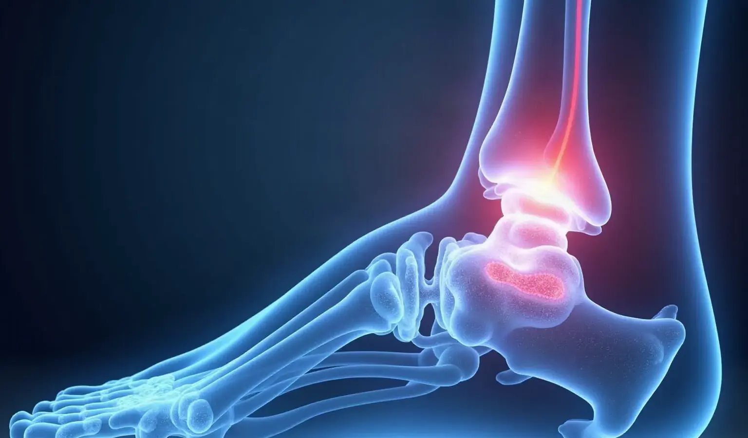
Inside-ankle pain is a symptom, not a diagnosis
Persistent pain along the inside of your ankle can make ordinary walking, exercise, and work feel uncertain. The location gives you a useful question to bring to an evaluation, but it does not identify the injured structure by itself.
A possible pattern involves the posterior tibial tendon, which runs along the inside of the ankle, attaches to the foot, helps turn the foot inward and point it downward, and helps maintain the arch. Pain in that area can also arise from other tendons, bones, joints, ligaments, or surrounding tissues. Use the pattern to organize what you notice—not to diagnose yourself.
That pattern is worth discussing when it persists, returns whenever you increase activity, or begins to affect how you use your foot.
A safe decision path has four stages:
- Identify the location and activity pattern without treating them as proof of a condition.
- Use cautious, supported care while you clarify what is happening.
- Move to professional assessment when pain persists, function changes, or an injury raises concern.
- Discuss rehabilitation, surgery, or regenerative options only after the clinical question and its limitations are clear.
The aim is not to select a treatment from a symptom description. The aim is to choose the next step that gives you better information without assuming a diagnosis.
Inside-ankle pain becomes more useful for decision-making when you describe what changes with movement, rest, and time. Record where the pain starts, which activity brings it on, whether the arch or ankle looks different, and whether you can use the foot normally. This record does not identify the cause, but it gives an ankle-focused clinician a clearer starting point.
Keep the first decision modest. You do not need to name the tendon, choose a procedure, or predict recovery before an assessment. You need to decide whether cautious supported care is reasonable for the moment, whether persistent symptoms justify an appointment, or whether an injury has made prompt evaluation more appropriate.
What the posterior tibial tendon does—and why its location matters
The posterior tibial tendon connects a muscle in the lower leg with the foot along the inside of the ankle. Its described actions include turning the foot inward, pointing the foot downward, and helping maintain the foot arch.
Those functions help explain why symptoms may become noticeable during walking, climbing, running, or other activities that load the ankle and arch. They do not tell you whether the tendon is inflamed, thickened, partially torn, or uninvolved. A similar location can accompany more than one problem, so the anatomy should frame your questions rather than settle the answer.
A case report illustrates why activity and function belong in the discussion. The report described a young female cross-country runner with stage two posterior tibial tendon dysfunction who reported pain rated 5/10 during running and 0/10 at rest. Her program included active rest, hip and ankle strengthening, manual therapy, and running-specific gait training.
That report describes one person and one treatment program. It does not show that the same stage, exercises, or outcome applies to you. Its practical lesson is narrower: explain what activity brings on your pain, what happens when you rest, and which movement or goal has become difficult. Those details can help a clinician decide what needs clarification.
Foot-arch support is one reason the posterior tibial tendon’s location matters during everyday movement. A tendon that helps maintain the arch is involved in a part of the foot that contributes to how you stand and walk, but pain near that structure does not show whether the tendon is the source of the problem.
Describe function in ordinary terms. Note whether you can walk on level ground, manage stairs, rise onto your toes, or return to your usual activity. These observations are more useful than trying to assign yourself a tendon stage from a diagram or symptom list.
Clues that make tendon involvement worth discussing
Activity-related changes are worth noting when you describe inside-ankle symptoms.
You may also notice changes around the ankle and arch. Possible signs include:
- Swelling, redness, or warmth along the inside of the ankle.
- A foot arch that appears flatter than before.
- Difficulty rising onto your toes.
- Pain that began gradually or has continued over several months.
These signs can make a clinical conversation more focused, but they cannot confirm posterior tibial tendon dysfunction. Do not use a flat-looking arch or difficulty with a heel raise as a self-assigned stage or severity label. Describe what you can and cannot do instead.
Reported factors associated with posterior tibial tendon dysfunction include a flatter foot shape, a tight Achilles tendon, unsuitable footwear, weak foot or leg muscles, and high body weight. These are reported risk factors, not a checklist that proves the tendon is responsible for your pain.
Your activity history also matters. Note whether symptoms follow a change in walking, running, stairs, uneven ground, footwear, or training load. Bring that account to an appointment, especially if you are an athlete, stay highly active, have diabetes, or have other foot-health concerns. Those circumstances can change the questions that need attention, but they do not supply a diagnosis on their own.
A short symptom record can separate a location clue from a function change. Write down:
- When the pain appears: at rest, during activity, afterward, or at more than one point.
- What happens after rest and whether ordinary walking becomes easier.
- Any visible swelling, warmth, redness, or change in the arch.
- Whether rising onto your toes is possible and how that movement feels.
- The activity, footwear, or surface that preceded a change.
Use the record to explain the pattern, not to grade the tendon yourself. A pattern that is gradual and activity-related can still need evaluation when it persists or affects function.

Start with cautious load reduction and supported conservative care
Conservative care for a possible posterior tibial tendon problem usually begins by reducing the activity that overloads the tendon while keeping the treatment question open. A cited review lists medial arch support and physical therapy focused on eccentric exercises among nonoperative options. A separate clinical leaflet lists reducing overloaded activity, progressive loading exercises, ice for pain reduction, supportive shoes, arch-support insoles, and a brace such as an Aircast with adjustable aircell for stage 1 dysfunction.
Those options belong to a general treatment discussion, not an individualized prescription. The right amount of activity, the need for a brace, and the timing of progressive loading depend on what is causing your pain and how your foot functions. If an exercise or support increases symptoms or changes your ability to walk, stop treating the plan as a fixed rule and arrange an assessment.
A cautious observation phase can help you decide whether you are improving, staying the same, or losing function. Keep the process simple:
- Reduce the specific activity that clearly aggravates the inside of your ankle.
- Avoid using temporary symptom relief as proof that the underlying problem has resolved.
- Notice whether ordinary walking, stairs, or rising onto your toes becomes easier, unchanged, or harder.
- Arrange professional evaluation when symptoms persist, recur with ordinary activity, or interfere with work, exercise, or daily movement.
Supportive footwear, arch support, bracing, and exercise should be discussed as parts of a plan rather than collected as disconnected fixes. A plan that accounts for your diagnosis, foot shape, current function, and activity goals gives you a clearer basis for deciding what to do next.
Conservative care should be judged by response and function, not by how many measures you can add. If reducing an aggravating load makes ordinary movement easier, that information can help shape the next discussion. If a brace, support, or exercise makes walking harder, that response is a reason to pause and seek guidance rather than increase the intensity.
Progressive loading is different from forcing painful activity. The cited treatment descriptions place loading within a broader plan that may include support, activity adjustment, and physical therapy. Keep the distinction clear: a general option is not a personal instruction, and improvement in one activity does not establish that the cause has been confirmed.
Why a gradual plan may be more realistic than a quick fix
Posterior tibialis tendon recovery may take time. A cited leaflet notes that the tendon may take a long time to heal because of its poor blood supply and states that there is no quick fix.
That limitation changes how you should judge a conservative plan. A single good day does not establish that the tendon has recovered, and a single painful day does not explain why symptoms returned. Look at function over time: can you walk, use stairs, or perform your usual activity with fewer limitations, or does the same load continue to reproduce the problem?
A gradual approach also protects you from treating discomfort as a race against a deadline. Do not increase activity simply because you want a faster answer, and do not continue a plan that is making function worse while waiting indefinitely. When the response is unclear, bring the pattern, activity changes, footwear, supports, and exercises you have tried to a clinician.
The practical boundary is straightforward: conservative care can be a reasonable treatment pathway when the situation is suitable, but persistent pain or declining function calls for clarification rather than repeated trial and error.
A useful progress check asks whether your activity tolerance is changing without treating time alone as proof of healing. Compare the same ordinary task across several observations: walking a familiar distance, using stairs, or standing long enough to complete a routine activity. Record whether symptoms appear sooner, later, or at the same point and whether your function changes afterward.
This kind of record cannot predict your recovery or establish the tendon’s condition. It can show whether the current plan deserves continuation, adjustment, or professional review. A gradual plan still needs a boundary.
Know when an injury changes the urgency
An ankle injury changes the question from gradual tendon irritation to possible structural injury. A fracture may cause swelling, pain immediately after the injury, bruising, inability to bear weight, or deformity. An X-ray is used to confirm whether a break is present.
If a fall, twist, impact, or other injury is followed by those changes, seek prompt medical assessment rather than relying on watchful self-care. Inability to bear weight or an ankle that looks out of place deserves urgent attention. The purpose is not to decide from symptoms whether you have a fracture; it is to avoid treating a potentially unstable injury as ordinary tendon soreness.
The injury pattern should also change what you tell the clinician. Give the time of the event, the movement or impact that caused it, whether swelling or bruising appeared immediately, and whether you can bear weight now. Do not force the ankle through activity to test it.
This section covers the supported injury warning signs that change the next step. It is not a complete list of every reason ankle pain may need urgent care. When the pain follows a significant injury and your ability to bear weight has changed, prompt evaluation takes priority over choosing among conservative or regenerative options.
A visible change in the ankle or an inability to bear weight is not a situation for testing different supports or waiting to see whether a regenerative discussion feels appropriate. Prompt assessment can determine whether an X-ray is needed and whether the injury requires a different pathway. Until you are assessed, avoid using activity to prove that the ankle is usable.
If the injury did not produce these changes but pain persists, the decision can return to the gradual pathway: describe the mechanism, track function, and arrange an ankle-focused evaluation when symptoms do not settle.
What an ankle-focused evaluation may need to clarify
An ankle-focused evaluation needs to connect your pain pattern with function and visible changes. A clinician may need to clarify whether the pain follows the posterior tibial tendon, whether swelling or warmth is present, whether the arch has changed, and whether you can rise onto your toes. Your activity history and any injury mechanism add context.
The same symptom location can lead to different treatment discussions, so the evaluation should answer a more useful question than “Which treatment should I try?” Ask what structure appears involved, what finding supports that view, what remains uncertain, and how your current function affects the plan. A symptom pattern can guide that conversation without becoming a diagnosis.
Treatment decisions also depend on how the tendon problem is classified and how you respond to nonoperative care.
That statement does not mean that three months automatically leads to surgery or that surgery is appropriate for every person with inside-ankle pain. It describes a review-based consideration tied to a diagnosed stage and an inadequate response to nonoperative treatment. If surgery enters the conversation, ask what finding makes it relevant, which nonoperative options have been tried, and what the proposed approach is intended to address.
Prepare for an evaluation with details that connect symptoms to use of the foot:
- The first day you noticed the pain and whether it began after an injury or gradually.
- The activities, surfaces, shoes, or training changes that affect it.
- Whether pain eases with rest or returns with walking and exercise.
- Any swelling, redness, warmth, arch change, or difficulty rising onto your toes.
- What supports, activity changes, exercises, or other care you have already tried.
The purpose of this preparation is not to arrive with a diagnosis. It is to give the clinician enough context to decide what needs examination and whether further assessment is relevant.
Imaging can inform the question without deciding it alone
Ultrasound and MRI do not produce one universal answer for every posterior tibial tendon question. Evidence comparing them is mixed because the studies used different populations, comparison methods, and diagnostic outcomes.
A study of 23 posterior tibial tendons in 23 patients with clinical findings of dysfunction found that high-resolution ultrasound and 3 T MRI were concordant in 20 of 23 cases, or 87 percent. A broader systematic review reported that MRI was more sensitive and specific overall for detecting posterior tibial tendon dysfunction. The same review reported ultrasound sensitivity of 80 percent and specificity of 90 percent for diagnosing tendinopathy, and sensitivity of 90 percent and specificity of 80 percent for diagnosing peritendinosis when compared with MRI. Sensitivity describes how often a test identifies a condition when it is present; specificity describes how often it is negative when that condition is absent.
The comparison can be kept in proportion:
| Imaging option | What the cited evidence found | Practical meaning for you |
|---|---|---|
| High-resolution ultrasound | One small study found agreement with 3 T MRI in 20 of 23 cases, or 87 percent, and slightly favored ultrasound in its comparison with operative findings. | This result comes from a small study and does not establish that ultrasound is the right test for every suspected problem. |
| MRI | A broader systematic review generally favored MRI for detecting posterior tibial tendon dysfunction. | MRI may be more useful for some detection questions, but test selection still depends on the suspected condition and the clinical assessment. |
Imaging should answer a defined clinical question rather than replace one. Ask what the clinician is looking for, how the result would change the plan, and whether the test fits the symptoms and functional changes you have described.
A test result also needs to be interpreted alongside your symptoms and examination. Agreement between two imaging methods in a small study does not mean either method settles every case, and a test characteristic reported in a review does not predict what your own result will show.
Before imaging, ask three practical questions:
- What specific structure or change is the test intended to assess?
- What would a positive or negative result change in the treatment discussion?
- Why is this test being considered for the symptoms and function you have described?
Those questions keep imaging connected to a decision instead of turning the scan itself into a diagnosis.

When conservative care has not been enough
Rehabilitation belongs in the treatment conversation even when the discussion turns to a procedure. Nonoperative options described for posterior tibial tendinopathy include arch support and physical therapy focused on eccentric exercises. A case report also described active rest, hip and ankle strengthening, manual therapy, and running-specific gait training for one runner with stage two dysfunction.
The case report does not establish a program for you. Its value is to show why treatment planning should connect the tendon question with strength, movement, activity demands, and the goal you want to regain. Ask how rehabilitation would fit the proposed pathway and what function the plan is intended to address.
Where regenerative treatment discussions fit
Regenerative treatment discussions can follow evaluation of the ankle problem rather than treating a symptom as a diagnosis. We offer clinical evaluations for patients considering regenerative treatment options for foot and ankle conditions. We personalize treatment plans around individual body and lifestyle needs.
That means a conversation can begin with your symptoms, activity goals, prior care, and the clinical question that still needs an answer. The treatment decision should remain tied to findings and to the limits of what is known.
Exosome therapy is one example of why careful wording matters. We describe exosome therapy as being investigated for its potential to support natural communication between cells during the body’s repair process, particularly in tendon and ligament tissue. Being investigated describes an area of interest; it does not establish that the approach will resolve your tendon problem.
We also explain that physiological responses to regenerative treatments vary significantly and that biological adaptation tends to follow a gradual timeline. Your discussion should therefore include what finding is being addressed, what uncertainty remains, how rehabilitation fits the plan, and how a response would be assessed over time. Regenerative care should be considered as an individualized clinical discussion, not as a promise of a quick fix.
The phrase regenerative treatment does not answer the questions that matter most for your ankle. You still need to know what structure is being considered, what finding supports the discussion, what established care has been tried, and what role rehabilitation would have. You also need a clear explanation that responses vary and that biological adaptation follows a gradual timeline.
Keep the boundaries visible. An option being investigated is not the same as an established answer for your symptoms, and an individualized plan is not a guarantee of resolution. A careful discussion can still be useful when it helps you compare that option with continued conservative care, further evaluation, or another treatment pathway.
What to ask before considering a regenerative option
Take these questions with you:
- What finding suggests that the posterior tibial tendon or another structure is involved?
- What has the examination clarified, and what remains uncertain?
- Which conservative measures are appropriate for my current function and activity goals?
- What role would rehabilitation play before, during, or after the proposed treatment?
- What evidence supports this option for the specific problem being discussed?
- What does “being investigated” mean for an option such as exosome therapy?
- How might physiological responses vary, and what time course should be discussed without promising an outcome?
A useful consultation should also leave room for a different next step. You may learn that continued conservative care, further assessment, or a surgical discussion is more relevant than a regenerative option. You may also learn that the available information does not yet identify a suitable treatment pathway.
The point of these questions is to connect the proposed treatment with your actual clinical findings. A personalized plan is meaningful only when you understand what is being treated, what is uncertain, and how progress or lack of progress would change the plan.
Before leaving the conversation, make sure you can explain the plan in plain language:
- The finding or clinical question being addressed.
- The conservative care that remains relevant.
- The role of rehabilitation and activity guidance.
- What uncertainty remains about the proposed option.
- How variable response and gradual biological adaptation affect expectations.
- What would lead to reassessment or a different treatment discussion.
If you cannot connect the proposed option to a finding and a practical plan, pause before treating the procedure as the next automatic step. The decision should remain understandable to you.
A safe next step for persistent inside-ankle pain
Persistent inside-ankle pain is best handled as a sequence rather than a single treatment choice. Use the location and activity pattern to frame a question about possible tendon involvement, not to diagnose yourself. When an appropriate plan is already being considered, supported care may include reducing overloaded activity, arch support, supportive footwear, progressive loading, physical therapy, or other measures selected for your situation.
If pain follows an injury with swelling, immediate pain, bruising, inability to bear weight, or deformity, seek prompt assessment because an X-ray may be needed to confirm a fracture. If symptoms continue, your ability to walk or rise onto your toes changes, or the cause remains unclear, arrange an ankle-focused clinical evaluation. For patients considering regenerative treatment, we offer clinical evaluations for foot and ankle conditions. A qualified clinician should decide whether any individual treatment is suitable for you.
The next conversation should clarify the finding, the conservative options, the role of rehabilitation, and the limits of any emerging treatment before you commit to a pathway.
Choose the lane that matches your situation: cautious supported care when symptoms are stable and no injury warning sign is present, prompt assessment when function or structure has changed, and a focused clinical discussion when persistent pain has not become clear. Keep the goal practical—understand what is being treated before choosing how to treat it.
Questions to ask before discussing regenerative treatment
How long should I try conservative care for inside ankle tendon pain before arranging an evaluation?
Arrange an evaluation when pain persists, returns with ordinary activity, interferes with walking or exercise, or function is not improving. Do not use a fixed time limit to diagnose yourself or decide on surgery. The cited review discusses surgery after three to six months of unsuccessful nonoperative care only when the condition and its stage have been clinically established.
Can I keep exercising with pain along the inside of my ankle?
Avoid or reduce the activity that clearly aggravates the pain rather than testing the ankle through worsening symptoms. General care may include supportive footwear, arch support, and gradual loading within an appropriate plan, but the right activity level depends on the cause and your function. If walking, stairs, or rising onto your toes becomes harder, seek clinical guidance.
What should I bring to an appointment for persistent inner-ankle pain?
Bring when the pain began, whether an injury or activity change preceded it, what movements worsen or ease it, and any swelling, warmth, redness, arch change, or difficulty rising onto your toes. Also list footwear, supports, exercises, activity changes, and other care you have tried. These details help clarify what needs examination without requiring you to arrive with a diagnosis.
What should I ask before considering regenerative treatment for an ankle tendon problem?
Ask what structure or finding is being treated, what remains uncertain, which conservative options and rehabilitation still matter, and what evidence applies to the specific problem. If an option is described as being investigated, ask what that means for your decision and how variable responses would be assessed. An individualized discussion does not guarantee that treatment is suitable or will resolve the pain.
Free consultation
Find out whether regenerative treatment is right for you.
Most consultations begin as a telehealth call with a physician specialist, so you can start without travelling to a clinic. No referral needed.